On the road to diffractive imaging of intrinsic molecular dynamics
09 September, 2015
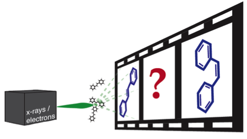
The determination of molecular structure and its changes over time is an essential step towards understanding chemical dynamics.
Time-resolved diffractive imaging is one promising approach to unravel structural changes, of nuclei and electrons, within a molecule. In the Controlled Molecule Imaging (CMI) group at CFEL two complementary diffractive imaging approaches, i.e., x-ray and electron diffraction, are employed to achieve a more complete picture of structural dynamics of gas-phase molecules.
Recently, two benchmark experiments have successfully been conducted and the results are published in special issues of the Journal of Physics B.
In the paper Strongly aligned gas-phase molecules at Free-Electron Lasers we present results on a highly efficient way to align molecules at Free-Electron-Laser (FEL) facilities. The alignment of molecules in the laboratory frame is advantageous over isotropic molecular samples, because not only bond length but also bond angles can be determined. This experiment was conducted at the Coherent X-ray Imaging (CXI) endstation of the Linac Coherent Light Source (LCLS) at the Sanford Linear Accelerator Center (SLAC). The molecular sample, 2,5-diiodothiophene, was aligned with intense laser pulses provided by the in-house Ti:Sapphire laser system. We demonstrate that it is possible to strongly align molecules with only a few mJ of pulse energy by utilizing the direct output of the regenerative amplifier on site. Thus we could record diffraction patterns, similar to previously obtained data at low repetition rate, but utilizing the full repetition rate, and thus all photons, of the FEL. We argue that the measured degree of alignment was limited by the intrinsic temperature of the molecule rather than by the alignment laser pulse. FEL facilities already have synchronized Ti:Sapphire laser systems, making our approach an easy implementation for future experiments.
Intense laser light, created by a Ti:Sapphire laser system, is also exploited for alignment at an existing setup in the CFEL lab of the Controlled Molecule Imaging group in Hamburg, in combination with a deflector to select molecules according to structure or quantum states — by exploiting strong and inhomogeneous electric fields. In the article Electron gun for diffraction experiments on controlled molecules, an electron gun was set up to perform diffraction experiments in a table-top setup. Controlling the molecules’ state and spatial orientation increases the amount of information contained in electron diffraction patterns. At the same time, the samples created by this apparatus are very dilute and laser alignment is only sustained in a very small interaction volume. Our recent contribution presents the newly set-up electron gun that addresses the requirements imposed by a dilute and small sample: The developed electron gun can produce up to 8 million electrons per pulse and uses an electrostatic lens for focusing to small enough spots. Moreover, the focusing electrodes are arranged in a configuration that can be used to measure the spatial and velocity distribution of the electron pulse at the cathode. In combination with simulations this allows for further characterization of the electron beam, as for example the determination of pulse duration and coherence length. As a final test of the suitability of the electron gun for diffraction experiments, diffraction data of a thin aluminum foil has been taken.
The electron gun is now combined with the controlled molecules setup and gas-phase diffraction data of controlled molecules can be recorded as the next step.
This approach, using electrons, will complement already conducted experiments with x-rays. Moreover, recent x-ray diffraction data, taken at the same set of experiments as in, is currently analyzed to determine the structure of 2,5-diiodothiophene.