Optically Controlled Particles for clean x-ray diffraction experiments
Approaches for the production of purified high-density beams of a broad variety of biological nanoparticles and establish control through electric fields and optical fields from a laser.
Laser induced alignment of nano-particles and biomolecules
Laser-induced alignment facilitates the retrieval of the 3D structure of single molecules imaged using x-ray free electron laser (XFEL) sources. It provides prior knowledge on the molecular orientation to the computer algorithms inverting the diffraction patterns to molecular structures. However, a strong alignment of nanoparticles and biological macromolecules in the gas phase has not been attained yet, and a search for the optimum experimental conditions to achieve the necessary degree of alignment of macromolecules is underway. A corresponding simulation framework would be highly beneficial. However, accurate modeling of laser-induced alignment through solving the time-dependent Schrödinger equation or even using the atomistic molecular dynamics (MD) is very challenging, if not impossible, for large molecules. Here, we present simulation results on the laser-induced alignment of gold nanoparticles and bio-macromolecules using classical rigid rotor dynamics.
The required molecular polarizability tensors are obtained through scaling of the tensors of perfect conductors of the same shape. We have also benchmarked different laser pulse profiles in order to optimize the achievable alignment and our simulations promising the necessary degrees of laser-induced alignment of nanoparticles and proteins for molecular-frame diffractive imaging.
Selection and control of (bio)nanoparticles with electric field
In single particle imaging (SPI) experiments, beams of individual nanoparticles are exposed to intense pulses
of x-rays from free-electron lasers (FEL) to record diffraction patterns of single, isolated molecules. The
reconstruction for structure determination relies on signal from many identical particles. Therefore, well defined-
sample delivery conditions are desired in order to achieve sample uniformity, including avoidance of
charge polydispersity.
Here, we present approaches for the production of purified high-density beams of a broad variety of biological nanoparticles. We establish control through electric fields, aiming at charge state or conformational state selectivity. This is especially relevant for soft biological samples, such as proteins or protein complexes, which in uncontrolled environment are prone to structural instability
Optical Injector of Particles for X-ray Diffractive Imaging
Motivated by the need for new single particle sample delivery methods to be used with our coherent diffractive imaging program we have developed an optical pipeline to produce a highly collimated stream of particles in either gaseous or vacuum environments. We present first results of our efforts to guide particles with micron-size precision in gaseous and vacuum environments using a first order quasi-Bessel beam operating either independently or collinear with a particle laden gas stream produced by an aerodynamic lens. The centimeter long, low divergence, optical pipeline is formed by a first order Laguerre–Gaussian beam imaged trough an axicon. When used in conjunction with an aerodynamic lens the optical forces assist the migration of particles to the centerline of the gas flow. This produces a higher particle number density and therefore an easier target to hit when probing with a Free Electron Laser (FEL) or other pulsed source. We present estimated optical forces exerted on the particles and the stiffness of trapping in the transverse plane, both depending on the particle size, optical reflectance, laser power, and background-gas pressure.
A 532 nm cw vortex beam was formed by a helical phase plate, and passed through an axicon with a base angle of 0.5° to form a first order Bessel like beam propagating over a meter-long distance. The slowly diverging beam was then re-imaged with a ×5 microscope objective into a narrow pipeline with minimum ring diameter of 2.4 μm and aspect ratio of approximately ×1000, which we termed as ‘optical syringe’ (Fig.1). 5-μm size spherical particles of graphite and sapphire, and 5-μm and 1.9-μm polystyrene spheres were used in our experiments to evaluate the optical force and scale it down to sub-micron particle size. We demonstrate that the resulting optical forces exerted on micron-size particles, using 5-W of laser power, were able to deflect the particle jet by about ~80 μm at the distance of 67 mm from the output nozzle of the aerodynamic lens stack.
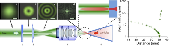
The experimental setup generating a micron-scale optical syringe based on an optical vortex combined with an axicon. (a-d) Images of the cross-sections of the beam before the phase plate (a), the 0.5° axicon (b), the microscope (c), and a magnified image of the beam near the nozzle of particle injector (d); the scale bar is 1 mm in (a-c) and 10 μm in (d). The inset illustrates the particle beam compression by optical forces. The graph on the right shows the beam radius vs. distance from the microscope.
Research Team
Lukas Haas, Muhamed Amin, Amit Samanta, Jochen Küpper
Andrei Rode (Australian National University, Canberra, Australia), Rick Kirian (Arizona State University, Tempe, AZ, USA), Henry N. Chapman (CFEL)
Acknowledgment
This work is supported by European Research Council under the European Union's Seventh Framework Programme (FP7/2007-2013) through the Consolidator Grant COMOTION (ERC-614507-Küpper) and by the excellence cluster “The Hamburg Centre for Ultrafast Imaging – Structure, Dynamics and Control of Matter at the Atomic Scale” of the Deutsche Forschungsgemeinschaft.